
Pin by Magpie on ชีวะ Prokaryotic cell, Eukaryotic cell, Prokaryotic cell model
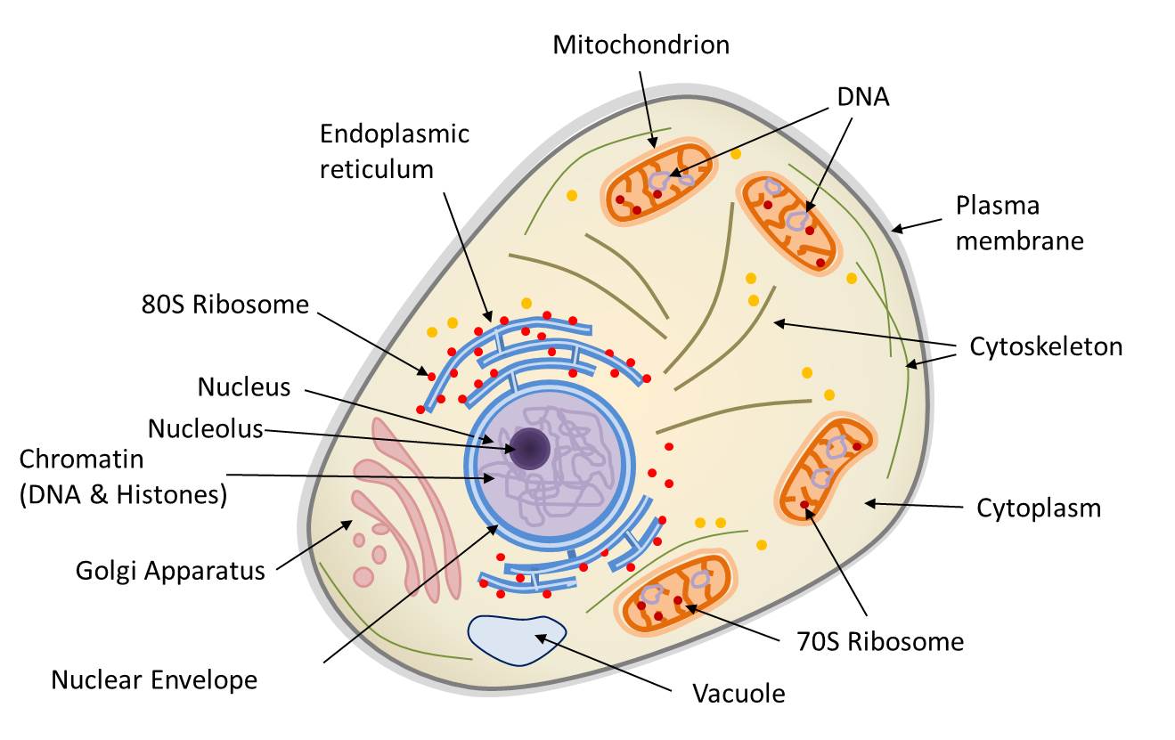
Two Types of Cells. There is another basic cell structure that is present in many but not all living cells: the nucleus. The nucleus of a cell is a structure in the cytoplasm that is surrounded by a membrane (the nuclear membrane) and contains, and protects, most of the cell's DNA. Based on whether they have a nucleus, there are two basic types of cells: prokaryotic cells and eukaryotic cells.

Draw a well labelled diagram of a prokaryotic cell Brainly.in
A prokaryotic cell is a type of cell that does not have a true nucleus or membrane-bound organelles. Organisms within the domains Bacteria and Archaea are based on the prokaryotic cell, while all other forms of life are eukaryotic. However, organisms with prokaryotic cells are very abundant and make up much of Earth's biomass.
Difference between Prokaryotic and Eukaryotic cell CBSE Class Notes Online Classnotes123
A prokaryotic cell is a type of cell that lacks a defined nucleus and other membrane-bound organelles. These cells are structurally simpler and smaller than their eukaryotic counterparts, the cells that make up fungi, plants, and animals. The genetic material in a prokaryotic cell is not housed within a nucleus; instead, it is contained in a.
Prokaryotic cell structure diagram, vector illustration cross section labeled scheme VectorMine
Prokaryotic cells are the unicellular cells that lack a well-defined nucleus, i.e. genetic material is not enclosed by a nuclear membrane. These cells are very minute in size 0.1 to 5.0 μ m. Common prokaryotic cell is a bacterial cell. Our body has over 100 trillion bacterial cells.

Simple Prokaryotic Cell Diagram Printable Diagram Cell wall, Biology, Microbiology
Eukaryotic cell: Prokaryotic cell: Size: Most are 5 μm - 100 μm: Most are 0.2 μm - 2.0 μm: Outer layers of cell: Cell membrane - surrounded by cell wall in plants and fungi:

3.258 imagens de Prokaryotes Imagens, fotos stock e vetores Shutterstock
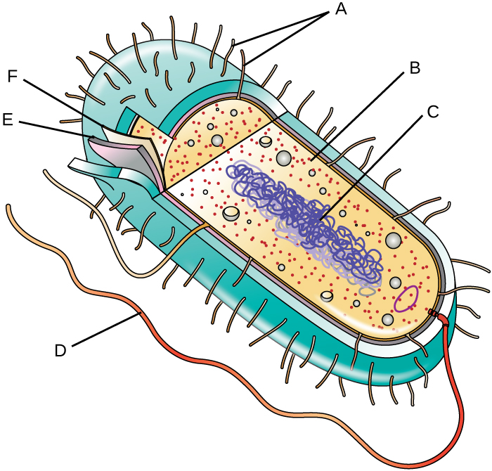
The main parts of a prokaryotic cell are shown in this diagram. The structure called a mesosome was once thought to be an organelle.. Can you identify each of the labeled structures?. The DNA of a prokaryotic cell is in the cytoplasm because the cell lacks a nucleus. Extracellular Structures. Many prokaryotes have an extra layer, called a.

Simple Prokaryotic Cell Diagram
Prokaryotic cell to label. Share Share by Susanmgillen. KS5 Biology. Show More. Edit Content. Embed. More. Leaderboard. Show more. Labelled diagram is an open-ended template. It does not generate scores for a leaderboard. Log in required. Theme. Fonts. Log in required. Options.
16 Important Differences between Prokaryotic and Eukaryotic cell CBSE Class Notes Online
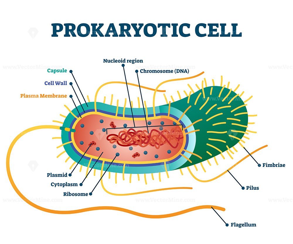
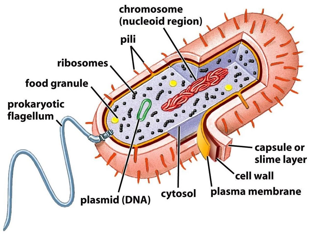
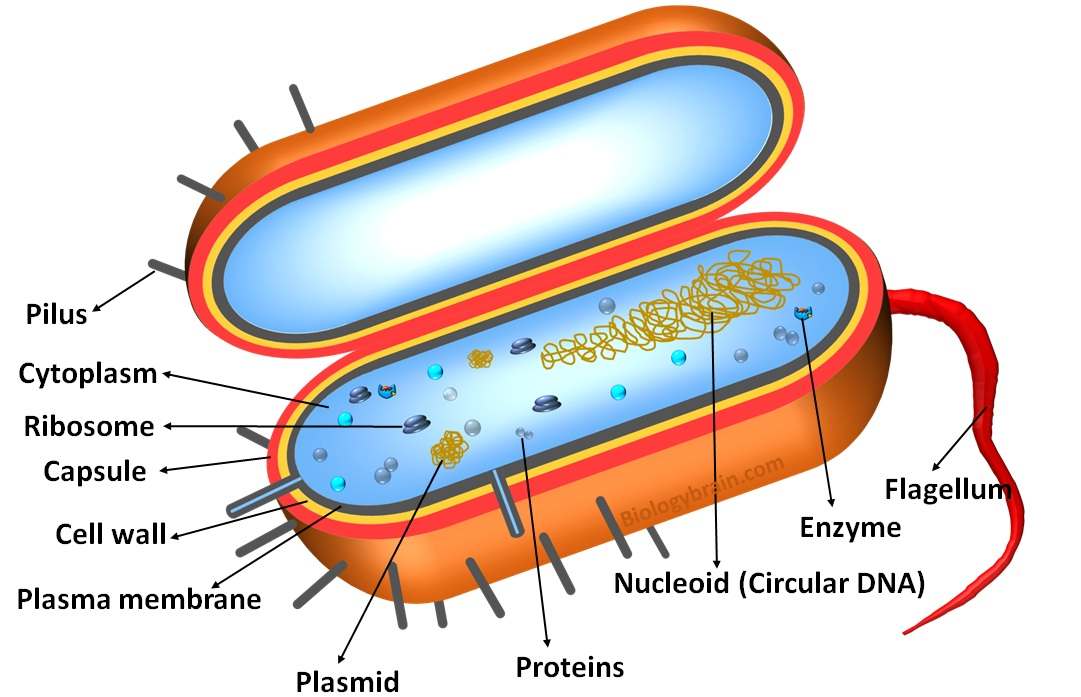
The Prokaryotic cell mainly has 4 components, that are: Ribosomes, Cytoplasm, Plasma Membrane, and DNA. Prokaryotic Cell Diagram. Here we have shown a well-labeled diagram of a prokaryotic cell. This diagrammatic representation of the prokaryotic cell will help you to understand the prokaryotic cell. Characteristics of Prokaryotic Cells

bacterial cell not labeled Clip Art Library
Prokaryotic cells are not as complex as eukaryotic cells. They have no true nucleus as the DNA is not contained within a membrane or separated from the rest of the cell, but is coiled up in a region of the cytoplasm called the nucleoid. Prokaryotic organisms have varying cell shapes. The most common bacteria shapes are spherical, rod-shaped.
Labeled Prokaryotic Cell Diagram, Definition, Parts and Function Biology Brain
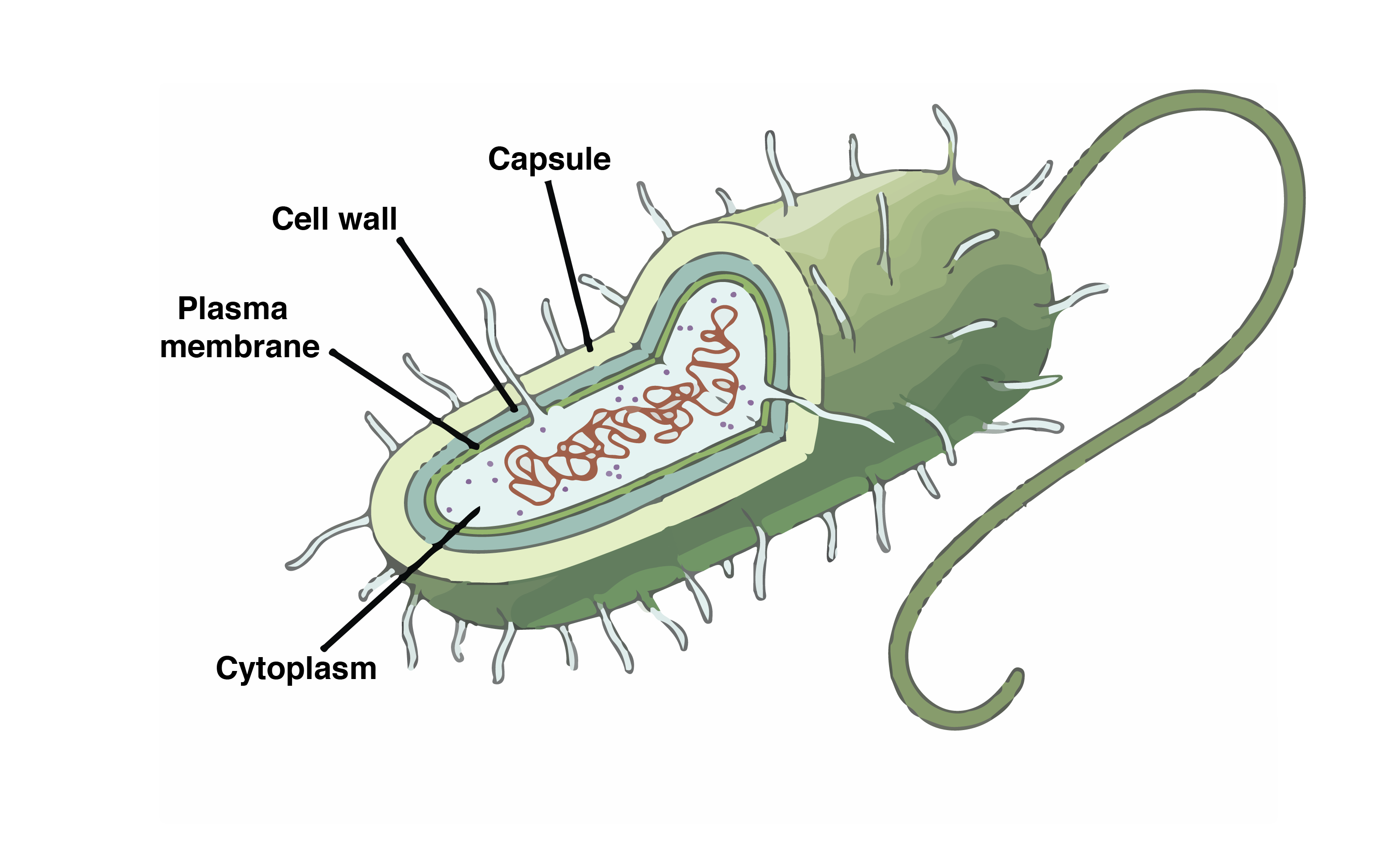
Key points: Prokaryotes are single-celled organisms belonging to the domains Bacteria and Archaea. Prokaryotic cells are much smaller than eukaryotic cells, have no nucleus, and lack organelles. All prokaryotic cells are encased by a cell wall. Many also have a capsule or slime layer made of polysaccharide.
IB Biology Notes Prokaryotic Cells Prokaryote Cell (Biology)
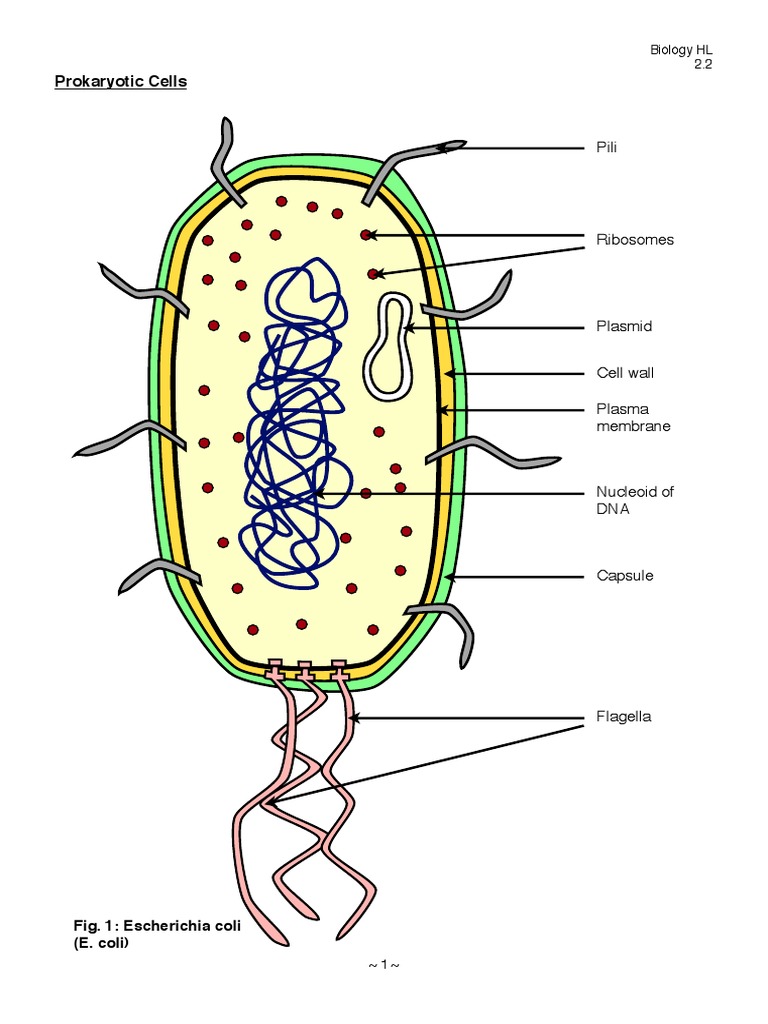
Prokaryotic cells 2.2.1 Draw and label a diagram of the ultrastructure of Escherichia coli (E. coli) as an example of a prokaryote.. 2.2.2 Annotate the diagram from 2.2.1 with the functions of each named structure.. Cell wall: Protects the cell from the outside environment and maintains the shape of the cell.It also prevents the cell from bursting if internal pressure rises.

Draw a well labelled diagram of a prokaryotic cell.
A well-labelled diagram of a prokaryotic cell consists of the following structures: Plasma membrane; Cell wall; Plasmid; Nucleoid (DNA) Pilus; Cytoplasm; Ribosomes; Capsule flagella; CONCLUSION. Our body acts as an ecosystem. The environment consists of both prokaryotic and eukaryotic cells and all the cellular structures can be categorised.

Prokaryotic Cell Diagrams 101 Diagrams
Animal and plant cells are types of eukaryotic cells, whereas bacteria are a type of prokaryote; Prokaryotic cells are much smaller than eukaryotic cells (between 100 - 1000 times smaller); They also differ from eukaryotic cells in having: A cytoplasm that lacks membrane-bound organelles; Their ribosomes are structurally smaller (70 S) in comparison to those found in eukaryotic cells (80 S)
36 Label The Structures Of The Prokaryotic Cell. Not All Terms Will Be Used. Labels 2021
Most prokaryotes have a cell wall outside the plasma membrane. Figure 27.2.2 27.2. 2: The features of a typical prokaryotic cell are shown. Recall that prokaryotes are divided into two different domains, Bacteria and Archaea, which together with Eukarya, comprise the three domains of life (Figure 27.2.3 27.2. 3 ).
3.3 Unique Characteristics of Prokaryotic Cells Microbiology Canadian Edition
cell membrane, cell wall, capsule/slime layer, plasmids, flagella, cytoplasm, circular DNA. 0%. prokaryotic cells. Share Share by Kross. KS4. Show More. Edit Content. Embed. More. Leaderboard. Show more Show less . This leaderboard is currently private.. Labelled diagram is an open-ended template. It does not generate scores for a.

Prokaryotic Cell Picture Labeled Anak Pak Lurah
1.11: Prokaryotic Cells. Distinguish between prokaryotic cells and eukaryotic cells in terms of structure, size, and the types of organisms that have these cell types. Identify structures of bacterial cells in models and diagrams, including details of Gram-positive and Gram-negative cell walls and flagella.